

If a surgeon has said the word microdiscectomy to you and you left the appointment without a clear picture of what actually happens, this article is for you. A microdiscectomy is an operation to remove the piece of disc material that is pressing on a nerve root in your spine. Micro refers to the small opening and the microscope, disc refers to the cushion between two vertebrae, and ectomy means taking something out.
What follows is a plain description of what happens during a microdiscectomy, step by step. It is a description, not a recommendation. Whether an operation is right for you depends on your imaging, your exam and your history, and that is a conversation for your own surgeon.
The quick version. A microdiscectomy removes the fragment of disc that is pressing on a nerve root, not the whole disc. It is done through a small opening in the lower back with a microscope or a magnifying loupe, and X-ray is used during the operation to confirm the right level. It is usually described as a decompression, because the point of it is taking pressure off a nerve.
What a microdiscectomy is
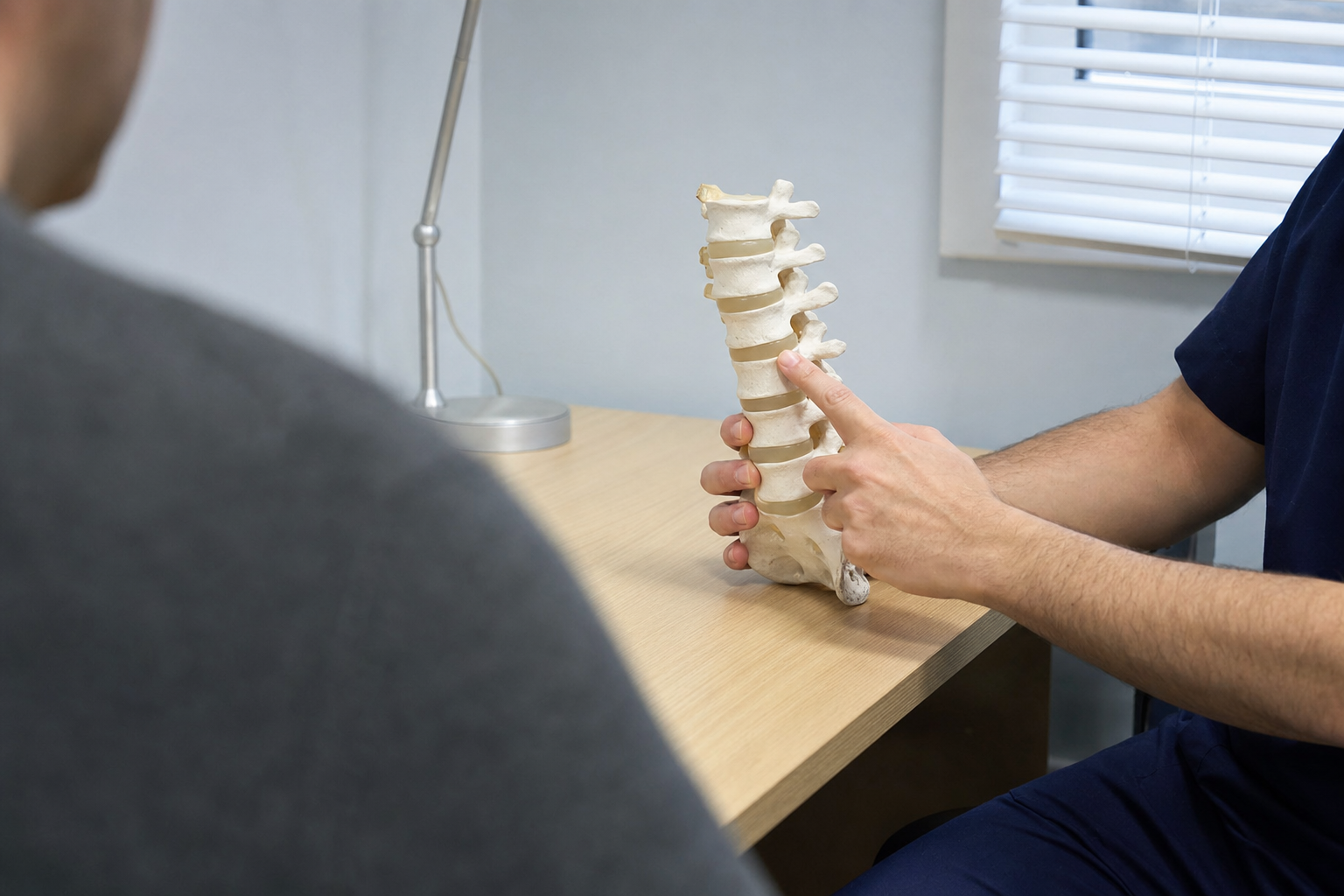
Between each pair of vertebrae sits a disc. Each disc has a tougher outer ring and a softer centre. When the outer ring tears, some of the softer material can push out through the gap. If that material lands against a nerve root, the nerve gets irritated and you feel it down the leg.
A microdiscectomy takes out the piece that is causing the pressure. The rest of the disc stays where it is. That surprises people, because the name sounds like the whole disc comes out. It does not. The surgeon is after the fragment and the small amount of loose material behind it.
Because the goal is taking pressure off the nerve, a microdiscectomy is grouped with the other decompression operations rather than with fusion. Nothing is fixed together and no hardware is put in during a straightforward microdiscectomy.
Why it comes up in a conversation about disc problems
Most people hear about a microdiscectomy after a herniated disc has already been found on an MRI and the leg symptoms have not settled with conservative care. Surgeons generally want to see that the imaging matches the exam, meaning the nerve root that looks compressed on the scan is the same one that explains the pain, the weakness or the changed sensation in the leg.
Discs also change with age, and that shows up on almost everyone’s scan eventually. Long Island Spine Specialists covers degenerative disc disease in detail, explaining the difference between normal wear on a scan and a disc problem that is causing symptoms. It is worth reading, because the two get mixed up a lot.
There are situations where a surgeon moves faster, such as significant weakness in a leg or changes in bladder and bowel function. Your surgeon will tell you if any of that applies to you. Everything else usually gets time first.
Getting ready on the day

The day itself is mostly waiting and checking. You arrive, change, and go through the same set of questions several times with different people. That repetition is on purpose. The team confirms your name, your procedure and the side and level being operated on more than once before anything starts.
An anesthesiologist talks to you about the anesthetic and your medical history. A nurse goes over the paperwork. Your surgeon usually comes to see you, marks the site, and answers whatever is still open. Then the surgical team scrubs and the room is set up while you are taken through.
A microdiscectomy is normally done under general anesthetic, so you are asleep for it. Your surgeon and anesthesiologist will explain what applies in your case, including how long you can expect to be in the hospital afterwards.
What the team does during the procedure

Once you are asleep, you are positioned face down with supports under the chest and hips so the lower back is accessible and the abdomen is free. The skin is cleaned and the area is draped. The photograph above shows a spine room set up in that way, with the imaging arm draped in clear plastic at the side of the table.
The table below sets out the usual order of the operation. Surgeons vary in the details and in the instruments they prefer, so treat this as the shape of it rather than a script.
| Stage | What happens | Why it is done that way |
|---|---|---|
| Confirming the level | An X-ray image is taken with a marker in place to identify the exact disc level | Lumbar levels look alike from the outside, so the level is confirmed with imaging rather than by eye |
| The opening | A small incision is made in the midline over that level and the muscle is moved to one side | Moving muscle aside rather than cutting through it is what keeps the opening small |
| Getting to the nerve | A small window is made in the bone or ligament at the back of the canal | This creates enough room to see the nerve root and work around it safely |
| Protecting the root | The nerve root is gently held to one side with a retractor | The disc fragment usually sits underneath or in front of the root |
| Removing the fragment | The loose disc material pressing on the nerve is taken out | Taking pressure off the nerve is the whole point of the operation |
| Checking and closing | The area is checked, irrigated, and the layers are closed with sutures | The surgeon confirms the root moves freely before finishing |
The removed material is small. People often expect something dramatic and are surprised when the surgeon describes a fragment the size of a fingernail clipping.
How it compares with other decompression surgery
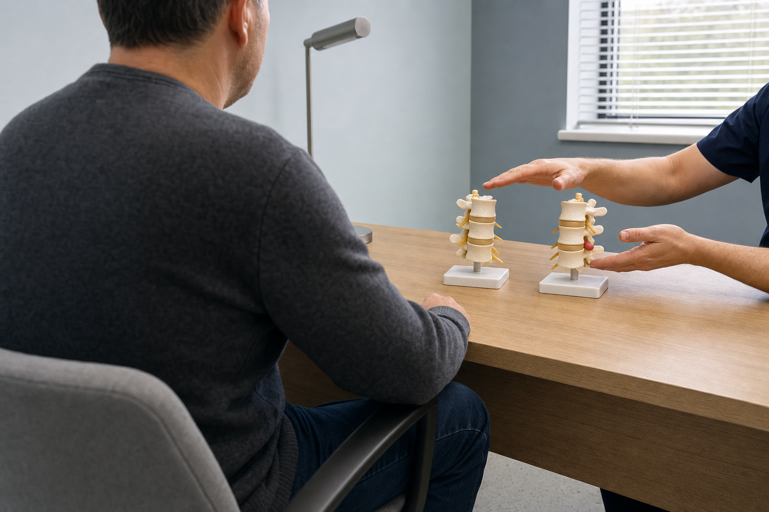
Microdiscectomy is one of several operations that take pressure off nerves. The names get used loosely, so it helps to know what each one describes. A laminectomy removes part or all of the lamina, which is the piece of bone at the back of the canal, and it is usually done when the canal itself has narrowed rather than when a single disc fragment is the problem.
| Operation | What is removed | Usual reason |
|---|---|---|
| Microdiscectomy | The disc fragment pressing on a nerve root | A herniated disc with leg symptoms that match the imaging |
| Laminectomy | Part or all of the lamina at the back of the canal | Narrowing of the spinal canal affecting the nerves inside it |
| Foraminotomy | Bone or tissue around the opening where a nerve exits | Narrowing at the exit point rather than in the main canal |
| Fusion | Nothing is removed to decompress; two or more vertebrae are joined | Instability or deformity, sometimes alongside a decompression |
A surgeon may combine these. It is common to hear a plan described with two names in it, and that is not a sign something has gone wrong. Ask your surgeon to draw it on paper if the words are not landing.
What the period after surgery generally involves
I am not going to give you a recovery timeline, because that belongs to the surgeon who did your operation and knows what they found. What I can describe is the shape of what usually gets discussed at the discharge conversation.
- Restrictions on bending, lifting and twisting, set by your surgeon and specific to your case
- How to look after the incision and what to watch for around it
- Walking, and how your surgeon wants you to build it up
- Whether physical therapy is part of the plan, and when it would start
- The symptoms that should prompt a phone call to the office rather than a wait
- When your follow-up appointment is and what will be checked at it
Write the answers down while you are still in the room. Most people remember about half of a discharge conversation, and the details you forget are usually the restrictions. Long Island Spine Specialists already has a longer microdiscectomy recovery guide that goes through the weeks afterwards, so I have kept this article on the operation itself.
Having the procedure done on Long Island
If you are weighing this up and want a spine-only practice to look at your case, Long Island Spine Specialists covers the whole range from conservative care through to surgery. They also offer second opinions, which matters if you have been told surgery is your only option and want another set of eyes on the imaging first.
The practice covers back surgery commack ny and minimally invasive spine surgery commack ny in detail, so you can read about what the Commack office handles before you call. Bring your MRI report and a written list of questions when you come in.
This article is general information about a procedure, and it is not a substitute for an assessment by a clinician who can examine you and review your images. Your surgeon is the person who can tell you whether any of it applies to your spine.
To arrange a consultation, use the contact form or call (631) 837-2359. Thanks for reading.