

If you have looked at yourself sideways in a mirror, noticed how much your lower back curves in, and wondered whether that lower back curve is normal or something called lordosis, this article is for you. I see this question a lot, usually from people who have just been told they have a curve and are not sure whether that was a diagnosis or a description.
This article explains what an assessment looks like and what a clinician measures. What your particular curve means is a question for the clinician who examines you and looks at your images. This article does not assess your particular curve or prescribe exercises.
The quick version. A curve in the lower back is normal and everybody has one. Lordosis is the anatomical name for that inward curve, so having lordosis is not the same as having a problem. The word only becomes a diagnosis when a clinician measures the curve, finds it well outside the usual range, and connects it to symptoms. That is done in person, not from a photograph.
Everyone has a lower back curve
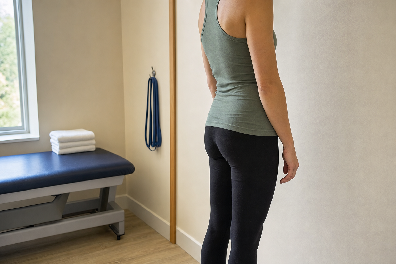
Look at a spine from the side and it is not a straight column. It curves forward at the neck, backward through the upper back, and forward again through the lower back. Those curves develop in childhood and they are there for a reason. They spread load, they let the spine absorb some shock, and they keep your head over your pelvis without you having to think about it.
So a hollow in the lower back is normal. The amount of it varies a lot between people. Body shape, muscle bulk, how someone stands when they are relaxed and how they stand when they know they are being looked at all change the picture. That is one reason a curve is measured rather than eyeballed.
It also changes through the day and through life. Pregnancy changes it. Carrying more weight around the middle changes it. Sitting for eight hours changes what it looks like when you stand up. None of that on its own is a condition.
What lordosis means as a word

Lordosis is the anatomical name for an inward curve of the spine. Your lower back has one and your neck has one. The opposite direction, the outward curve through the upper back, is called kyphosis. Both words describe shape, not damage.
This is where the confusion starts. If a report says lumbar lordosis, that is often just naming the normal curve. If someone says you have lordosis in a worried tone, they usually mean the curve is more pronounced than average. The word is doing two jobs and the tone is carrying the difference, which is not a great system.
If you want the two curve types laid out side by side, Long Island Spine Specialists has a write-up comparing kyphosis and lordosis that goes through both in detail. It is a useful read before an appointment, because it gets the vocabulary straight.
When a curve gets called too much or too little

Clinicians use a few different terms depending on which direction a curve has gone and how far. Here is what the common ones actually describe.
| Term | What it describes | How it is normally assessed |
|---|---|---|
| Lumbar lordosis | The normal inward curve of the lower back | Observed on a standing exam and measured on a standing X-ray if needed |
| Hyperlordosis | An inward curve that is more pronounced than the usual range | Measured on imaging and considered next to the exam findings |
| Hypolordosis or flatback | A lower back that has lost some of its normal inward curve | Same approach, looking at the angle from the side |
| Kyphosis | The outward curve of the upper back, or an increased version of it | Assessed from the side, often at the same appointment |
| Scoliosis | A sideways curve and rotation, which is a different plane entirely | Assessed from behind as well as from the side |
The practice has a separate write-up on hyperlordosis covering causes, symptoms and how it gets treated, so I am not going to repeat that ground here. What I want to get across is that the label comes from a measurement plus an examination, not from how your back looks in a photograph.
What a clinician looks at when you stand

A standing assessment is usually the first thing that happens, and it is quick. The clinician looks at you from the side and from behind, in normal standing rather than a corrected posture. They are watching where your head sits over your ribs, where your ribs sit over your pelvis, and whether the two sides look level.
Then they usually run their fingers down the line of the spine to feel the bony landmarks, because what the skin does and what the bones do are not always the same thing. They will look at how the pelvis is tipped, since a pelvis rotated forward makes the lower back curve look deeper without the spine itself having changed.
Movement comes next. You will probably be asked to bend forward, lean back and side bend, while the clinician watches whether the curve reverses smoothly or stays locked in one shape. They are looking at how the curve behaves, not just what it measures at rest.
The rest of the assessment, and what imaging adds
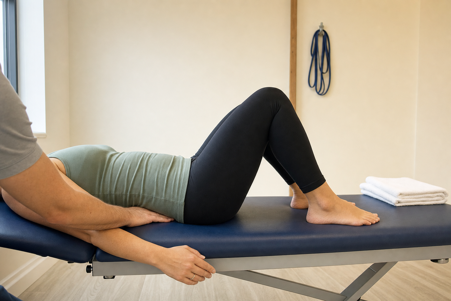
After the standing part, most assessments move to the treatment table. Lying down takes gravity out of it, and the difference between how a curve looks standing and how it behaves lying down tells a clinician something. Hip range of movement usually gets checked at the same appointment, because the hips and the lower back move together.
Imaging is where a curve stops being a description and becomes a number. A standing X-ray from the side lets a physician measure the angle between specific vertebrae, and that measurement is what gets compared against the usual range. An MRI is a different job and shows the discs, the nerves and the soft tissue rather than the alignment.
That review happens in person. Long Island Spine Specialists sees patients at its Commack office among others, and that office is the practice’s orthopedic commack location where a physician can sit down with your images and go through them with you. If you want to know who you would be seeing, there is a profile for the spine specialist commack patients book with there.
What else gets looked at alongside the curve
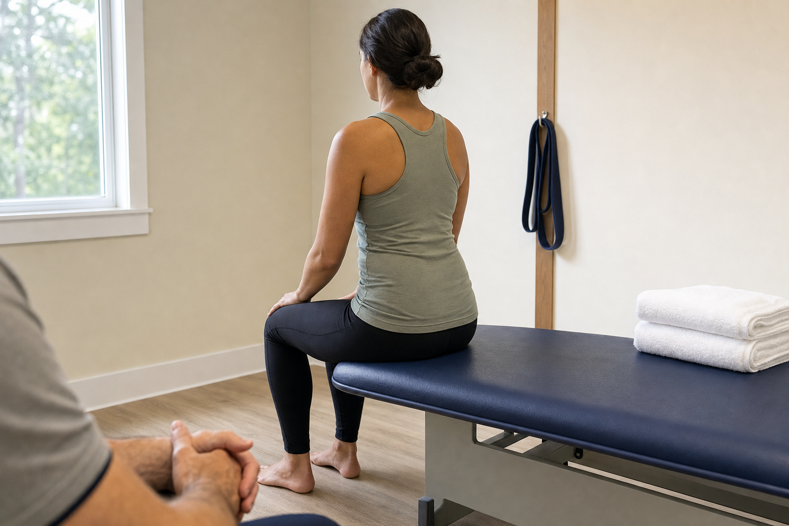
A curve on its own is rarely the whole story. When someone comes in about their lower back shape, a clinician is usually thinking about a short list of other things at the same time.
- Whether there is any pain at all, and where it sits when there is
- Whether symptoms travel into the legs, which points the assessment in a different direction
- Hip mobility and how the pelvis sits, since both change what the curve looks like
- Whether the curve is flexible or fixed when you move
- Anything in the history such as a previous fracture, surgery or a known condition
- How long the shape has been there, and whether anyone has measured it before
Some of those answers open other conversations. A deep curve with leg symptoms that come on when walking, for example, would have a clinician thinking about spinal stenosis rather than the curve by itself. Where the main complaint is the back rather than the shape, the practice’s section on back pain relief covers the range of what gets offered, from conservative care through to surgery.
Getting your curve looked at on Long Island
If the shape of your lower back has changed, or it has been there a long time and now something hurts, the useful move is having it measured properly rather than comparing yourself against pictures online. An assessment takes one appointment and it either gives you a number or it tells you the curve was never the issue.
Long Island Spine Specialists is a spine-only practice with offices across Long Island, and it works alongside physical therapists and chiropractors rather than replacing them. If you already have a therapist you like, bring their notes. Bring any previous imaging as well, because a comparison over time is worth more than a single picture.
This is general information about how curves are assessed and is not a substitute for an assessment by a clinician who can examine you. Questions about measurement and diagnosis should be discussed with that clinician.
You can request an appointment through the contact form or call (631) 837-2359. Thanks for reading.